Showing posts with label adipocytes. Show all posts

Monday, November 14, 2011

Weight loss in a pill


Source: Wikipedia.  Butter (fat) melting away...


What if you could take a pill that would simply melt your fat away?  The dream has become reality, but you have to be a chubby monkey to get your hands on it.

Barnhart and colleagues performed a placebo-controlled trial on adipotide, an experimental new drug to combat obesity, in obese rhesus monkeys.  Adipotide acts to slowly destroy the blood vessels that feed fat tissue.  By starving fat cells nutrients and oxygen, the cells eventually die, and fat loss ensues.  The compound has shown considerable success in rodents, but in order for the drug to progress into human trials, the experiments had to be replicated in non-human primates.  Indeed, the results are promising.

Spontaneously obese monkeys were given daily injections of adipotide, or placebo, for 28 days, followed by a 28 day recovery period.  The treatment group enjoyed a 15% weight loss, on average, by the end of the eight weeks – equivalent to a 275 pound person losing 40 pounds.  The bulk of the fat loss was visceral fat, which is the most deleterious region to carry body fat.

There is still plenty of time until it could be use to treat human obesity, but until then, the drug’s actions on fat tissue are interesting to ponder.
Souce:  Wikipedia.  Adipose (fat) tissue. 
The yellow fat cells are usually supplied by the red blood vessels.


Adipotide is a protein-based compound that binds to the endothelial cells that line the vasculature of fat tissue.  The protein enters the cells and causes them to commit planned suicide, or “apoptosis” in biology-speak.  Deprived of blood, the fat cells progressively die-off as they starve of oxygen and nutrients.  The fatty acids and triglycerides within the cells are released into the circulation and the body cleans-up the debris, leaving the monkeys svelte.  But where does the fat go?

One would expect a flood of triglycerides into the blood stream.  This is called hypertriglyceridemia and is a component of the metabolic syndrome and is a risk factor for heart disease.  However, this doesn’t appear to be the case.  The animals’ blood lipids improved throughout their weight loss.  What’s more, the animals became considerably more insulin sensitive – an important improvement that prevents diabetes and other complications of obesity.  Together, these data suggest that the freed fat was successfully oxidized, or “burned”, by the body for energy.

Throughout the treatment period, the monkeys receiving adipotide consumed fewer “biscuits” than their overweight controls.  The authors attribute the poor appetite to enhanced satiety, rather than nausea, which in turn led to weight loss.

However, adipotide did not cause lean monkeys to eat less.  The selective effect on appetite suggests that eating less isn’t the primary effect.  And after all, the drug targets the fat cells, not the brain.  Rather, it seems plausible that the dying fat cells free up an abundance of fat to be used for fuel.  With plenty of fuel available to the body, there’s little reason to stock up on biscuits.

There isn’t any data measuring the type of fuel – carbohydrate or fat – metabolized by the monkeys, so the mechanism requires a bit of speculation. 

The monkeys lose fat mass into the circulation, which gets oxidized for energy.  Fat loss improves insulin sensitivity, so the animals don’t have to secrete as much insulin in order to compensate for insulin resistance.  Plenty of fat for energy in the presence of lower insulin – remember, insulin drives fat storage – allows the monkeys to get by with eating less.  It’s like a drug-induced low carb diet, but instead of a bun-less cheeseburger, the monkeys are able to dine on their fat stores.  Although the cheeseburger would have one less side effect.

Source: Wikipedia.  A lean (and wild) rhesus macaque


The experimental monkeys showed transient problems with their kidneys, as indicated by elevated creatnine levels and slight microscopic damage to the tissue.  It’s unclear from this study whether the problem stems from the drug itself or a complication from the dying fat cells.  Side effects are especially problematic if adipotide needs to be taken chronically.

The monkeys started to show some regain of weight by the end of the four week recovery period, but the amount was minimal.  This could be an inherent problem to the drug, or it could be because they stuck to their low-fat junk food diet - monkey chow composed of 59% carbohydrate, 28% protein, and 12% fat – that made them fat in the first place.

Thursday, October 6, 2011

A molecular biologist serving pizza and fatty liver


Source:  Wikipedia.  I would have had some if it looked this good...

There’s few things more ironic than walking into a lecture titled “the molecular biology of hepatic steatosis” and being met by a table stacked with pizza and soda.  Of course, the molecular biologists in the crowd weren’t the ones with non-alcoholic fatty-liver disease (or NAFLD), but it’s hard to ignore the dissonance.  Fortunately, the talk was better than the lunch offerings.

The lecture was by a medical researcher who investigates the molecular mechanisms behind fatty liver disease.  Today he was highlighting his group’s most recent work on the molecular mechanisms connecting obesity to liver fat deposition.

Source: Wikipedia.  Adipocytes (fat cells).

The researcher wanted to address several hypotheses.  The first hypothesis is that when people become obese, their fat cells enlarge (rather than multiply) to a point that induces cellular stress.  This stress then produces a cascade of intracellular signals that tell the fat cell (adipocyte) to begin apoptosis (intentional cell death).  The troubled fat tissue then secretes deleterious cytokines, or hormone-like chemicals.

Source:  Wikipedia.  A Macrophage.

The second hypothesis is that these cytokines recruit immune cells – specifically macrophages, an important cell of the innate immune system – that begin to engulf the fat cells.  The combination of dying fat cells and macrophages causes a big problem.

When fat cells become too large, and start dying off, they dump their fat content into the circulation – like an overstuffed cream-filled donut.  This results in a surplus of free fatty acids floating around the body.  And this is important because two-thirds of the fat in the livers of people with NAFLD are derived from the circulation.  And it isn’t just the adipocytes causing trouble.

Macrophages enjoy company, so they recruit more macrophages by secreting cytokines that have wonderful names such as Tumor Necrosis Factor alpha and Interleukin-6.  These chemicals, and many others, create a vicious cycle whereby inflammation produces more inflammation produces more information.  The inflammation in the fat then appears to spill out into the circulation and reach the liver.

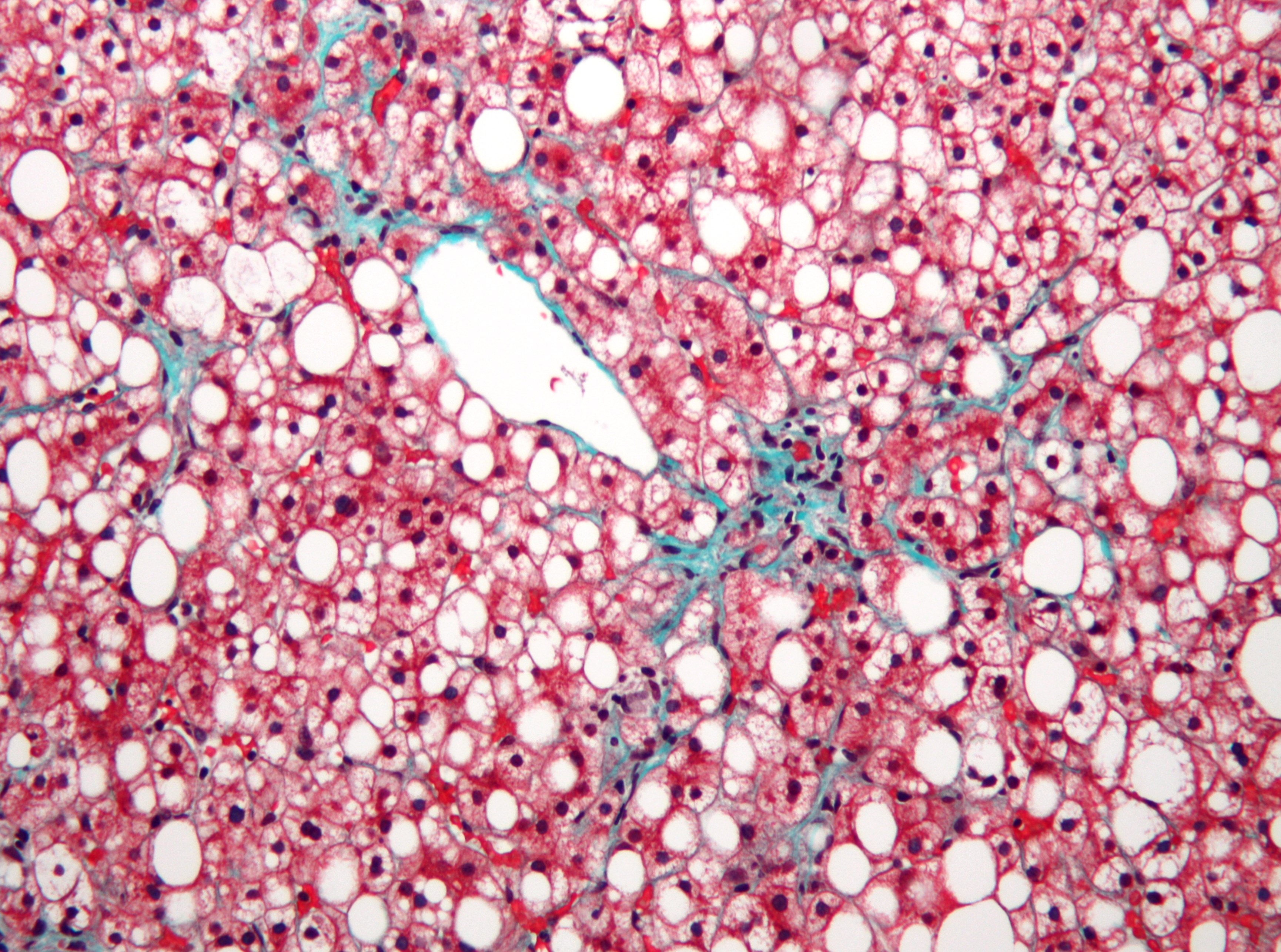
Source: Wikipedia.  Not-alcoholic fatty liver disease.
Liver cells are pink,  the white is fat that shouldn't be there...

Inflammation in the liver leads to dysfunctional fat metabolism.  The liver then begins producing too much fat, which also accumulates in the liver.  The excessive free fatty acids in the circulation and the dysfunctional fat metabolism in the liver account for virtually all of the excess fat seen in NAFLD.  This fat then begets more inflammation.

The excessive fat and inflammation in the liver, and the fat from inflammation, generate a lot of oxidative stress in the liver.  This oxidative stress produces more inflammation, and causes the liver cells to dye off.  This can lead to a clinically inflamed liver (steatosis hepatitis) and even cirrhosis (think alcoholic).  At least, according to the researcher's hypothesis.

The researcher had plenty of data from cell cultures and mice, each experiment clearly showing an increase in relevant proteins and genes in response to diet-induced NAFLD.  He also cited a clinical trial that showed that vitamin E (an anti-oxidant) supplementation was more beneficial than the insulin-sensitizing drug metformin in patients with confirmed NAFLD.  Although, admittedly, the anti-oxidant treatment didn’t seem to be that much of a better treatment.  But perhaps oxidation is indeed the culprit, and Vitamin E just isn’t a strong enough anti-oxidant. 

The obesity to inflammation to fatty liver (and other problems) is a compelling hypothesis, and it has plenty of support in the research community.  If this hypothesis with stands the test of time, then it would support the notion that whatever makes us fat (sugar ‘cough’ and ‘cough’  white flour), probably also leads to insulin resistance, fatty liver, and all the other diseases associated with the Western diet.  But however it goes, I’m still glad that I brought tuna, broccoli, and buttered brown rice instead of chowing down on cheap pizza and soda.

Thursday, May 5, 2011

Fat cells: Is it the size or the number?

Obesity is tightly associated with virtually all "western diseases," including high blood pressure, diabetes, gout, and dyslipidemia.  Insulin resistance (type 2 diabetes) is similarly associated with these diseases.  This is why researchers have begun to use the literary gem and public health nightmare "diabesity" when referring to our current health epidemic.  Thus, researchers are diligently trying to tease apart the associations and mechanisms that explain diabesity.
  
Veilleux and colleagues published a study in this month's issue of Diabetes looking at the predictive power of the size and number of adipocytes (fat cells) on markers of dyslipidemia ("poor blood lipids").     They found that the enlargement of abdominal adipocytes was independently associated with changes in blood lipids consistent with the metabolic syndrome.

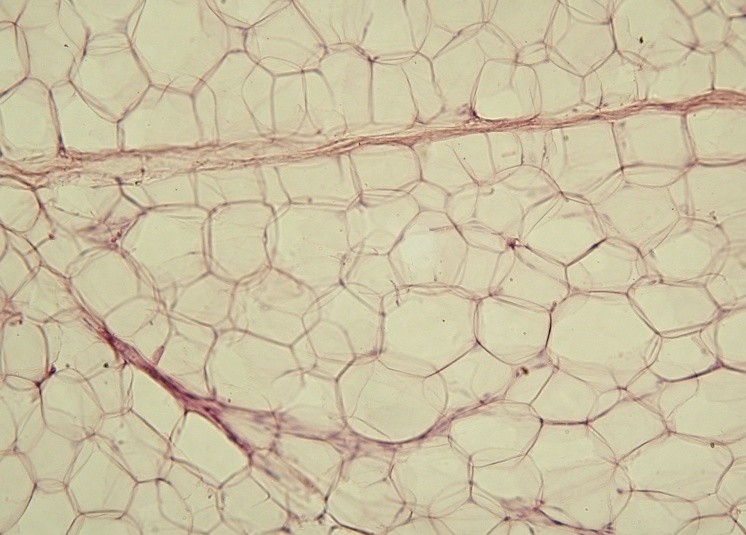
Source: flickr
Above is a photomicrograph of adipocytes.  The dark splotches are the nuclei of the cells.  The big white space?  That's a large lipid droplet.  Adipocytes store fat to be used for energy, and as you can see, they are very good at what they do.  Our fat stores can increase by either enlargement of the adipoctyes or an increase in the number of adipocytes, or as is common in biology, a combination of the two. And all of these cells aggregrate to form different fat depots in the body.  
    
Two of the main fat stores are subcutaneous fat - meaning under the skin - and omental fat, which rests behind the abdominal wall.  The gynoid, or "pear shape," fat distribution refers to subcutaneous fat distributed around the hips and buttucks, and it is associated with a decreased risk of dyslipidemia.  Android, or "apple shape," fat distribution refers to abdominal obesity, and is associated with an increased risk of dyslipidemia.  But remember, a large gut is composed of both subcutaneous fat and omental fat; both tend to increase together but it is the omental that seems the most deleterious.
Abdominal subcutaneous fat
Source: FreeDigitalPhotos

Peripheral subcutaneous fat
Source: FreeDigitalPhotos

Given the known complications of body fat, researchers wanted to look to see if the association between omental fat and dyslipidemia was more closely related to enlarged adipocytes or a higher number of adipocytes, independent of fat distribution or fat mass.  Obese women who were undergoing surgery had biopsies taken of their omental and subcutaneous fat.  These biopsies were then used to categorize the women by adipocyte hypertrophy (enlargement) or hyperplasia (high cell count) for both subcutaneous and omental fat stores.  Blood markers for dyslipidemia - particularly representing hypertriglyceridemia, or elevated triglycerides, were then coupled with the adipocyte data to develop a predictive model - that is, how do adipocyte characteristics predict dyslipidemia.

The most pronounced finding was that sufficiently large omental adipocytes increased the risk of hypertriglyceridemia by 4-fold, whereas sufficiently many omental adipocytes only increased the risk by 1.5-fold.  The size or number of subcutaneous adipocytes showed no increased risk of elevated triglycerides.  These changes in risk were seen after several factors had been adjusted for - such as body fat mass, BMI, and others - and so represent the independent contribution of these adipocyte characteristics.  The enlarged omental adipocyte group further demonstrated dyslipidemia in other triglyceride-related blood markers, although the results were less dramatic.

Figure: Conceptual representation of the results
And perhaps you are wondering, do enlarged omental adipocytes cause the dyslipidemia?  Does the dyslipidemia cause the enlarged adipocytes?  Or is there something else that causes them both to occur in lockstep?  An interesting piece to the puzzle is that surgical removal of omental fat does not improve dyslipidemia.  Or only seems to do so in the short-term when it is accompanied with the already-shown-to-do-so gastric bypass surgery (article on recent findings).  Those data would indicate that it is not the omental fat that causes the dyslipidemia.  But given the Veilleux study we just discussed, perhaps surgical removal would work if the patients had predominately large omental adipocytes, rather than many.  

While it's well known that obesity is tightly associated with dyslipidemia and metabolic syndrome, these data are the first to specifically examine adipocyte "cellularity" with metabolic derangement in obese women.  Admittedly, this experiment is more interesting from a physiological or mechanistic standpoint - everyone knows that abdominal obesity is both unhealthy and no one is happy with their "gut" regardless of the cellular mechanism.  So while we wait for that eventual discovery, let's agree to keep our visceral fat cells both small and few.